
Exercise 9 the axial skeleton – Embark on an enlightening journey with Exercise 9: The Axial Skeleton, where we delve into the intricate world of bones that form the core of our body. Brace yourself for a captivating exploration of the skull, vertebral column, and rib cage, revealing their remarkable functions and interconnectedness.
From protecting vital organs to facilitating movement, the axial skeleton plays a pivotal role in our overall well-being. Join us as we unravel the secrets of this fascinating skeletal structure, leaving you with a profound appreciation for its complexity and significance.
The Axial Skeleton
The axial skeleton is the central axis of the skeletal system, providing support, protection, and mobility for the body. It consists of the skull, vertebral column, and rib cage, which together form a rigid framework that houses and protects vital organs, facilitates movement, and allows for various body functions.
The axial skeleton’s primary function is to protect and support the head, neck, and trunk. It provides a bony framework that encloses and safeguards the brain, spinal cord, and internal organs, such as the heart and lungs. Additionally, the axial skeleton plays a crucial role in movement, allowing for flexion, extension, rotation, and other movements of the head, neck, and trunk.
The Skull
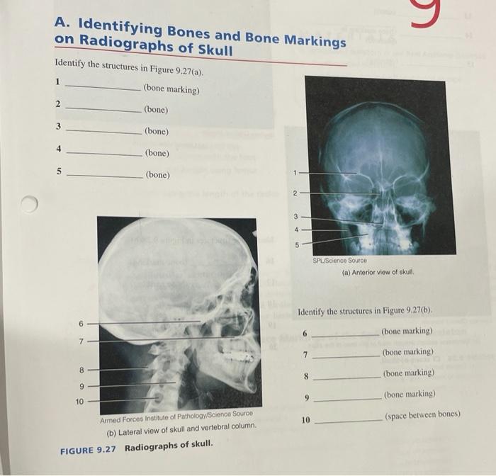
The skull is the bony structure that encloses the brain and provides protection for the facial structures. It consists of 22 bones that are tightly connected to form a rigid and protective case. The skull can be divided into two main parts: the cranium and the facial bones.
The cranium, or braincase, is the upper and larger part of the skull that houses and protects the brain. It consists of eight bones that are fused together to form a strong and solid structure. The facial bones are the lower and smaller part of the skull that forms the face.
They include the bones of the eye sockets, nose, and jaw, which provide support and shape to the face.
The Vertebral Column
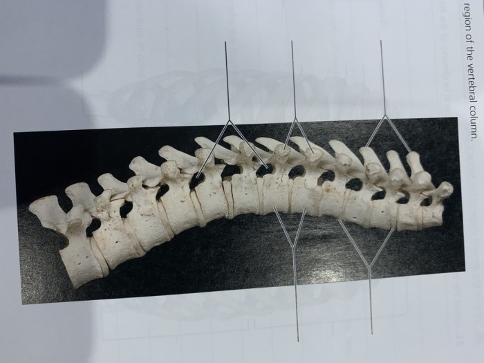
The vertebral column, also known as the spine or backbone, is a flexible and segmented bony structure that extends from the skull to the pelvis. It consists of 33 vertebrae, which are stacked one on top of the other to form a protective canal that houses and protects the spinal cord.
The vertebrae are interconnected by ligaments and muscles, allowing for a wide range of movements, including flexion, extension, rotation, and lateral bending.
The vertebral column is divided into five regions: cervical (neck), thoracic (chest), lumbar (lower back), sacral (pelvis), and coccygeal (tailbone). Each region consists of a specific number of vertebrae that are adapted to perform different functions and provide support to different parts of the body.
The Rib Cage
The rib cage is a bony cage that encloses and protects the thoracic cavity, which contains the heart, lungs, and other vital organs. It consists of 12 pairs of ribs that are attached to the thoracic vertebrae and the sternum (breastbone) at the front of the chest.
The ribs are flexible and allow for expansion and contraction of the chest during breathing, facilitating the exchange of gases in the lungs.
The rib cage plays a crucial role in respiration and provides protection for the internal organs. It also serves as a point of attachment for muscles that are involved in breathing and other movements of the trunk.
Functions of the Axial Skeleton: Exercise 9 The Axial Skeleton
The axial skeleton, comprising the skull, vertebral column, and rib cage, serves multifaceted roles in supporting, protecting, and facilitating movement within the body.
Protective Function
The axial skeleton’s primary function is to provide protection for vital organs and structures. The skull, composed of a series of fused bones, encases the delicate brain, shielding it from physical impact and external damage. Similarly, the vertebral column, consisting of a series of interlocking vertebrae, encloses the spinal cord, a critical communication pathway between the brain and the rest of the body.
Support and Stability
The axial skeleton provides structural support for the body, enabling it to maintain an upright posture and withstand external forces. The vertebral column, with its curved shape and interlocking vertebrae, acts as a central pillar, distributing weight and maintaining balance.
The rib cage, formed by the ribs and sternum, creates a protective cavity for the heart, lungs, and other vital organs.
Exercise 9: The Axial Skeleton is a comprehensive guide to the bones of the head, neck, and trunk. For those seeking further practice, abeka business math test 10 provides a thorough review of business mathematics concepts. Returning to Exercise 9: The Axial Skeleton, students will delve deeper into the structure and function of these vital skeletal components.
Movement
The axial skeleton also plays a role in facilitating movement. The skull allows for head rotations, enabling us to change our field of vision and respond to stimuli. The vertebral column, with its flexible joints, allows for bending, twisting, and other complex movements of the neck, back, and trunk.
The Skull
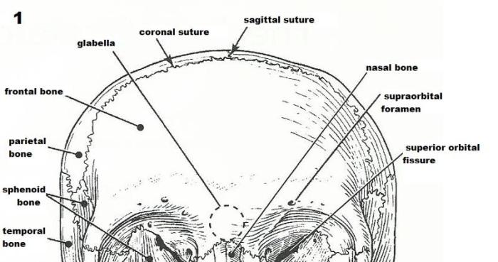
The skull, a complex and fascinating structure, forms the skeletal framework of the head and serves as the primary protective casing for the brain. It comprises a total of 22 bones, meticulously fused together by intricate joints called sutures. These sutures allow for slight movement during birth and growth, but eventually fuse into solid, immovable connections in adulthood.
Bones of the Skull
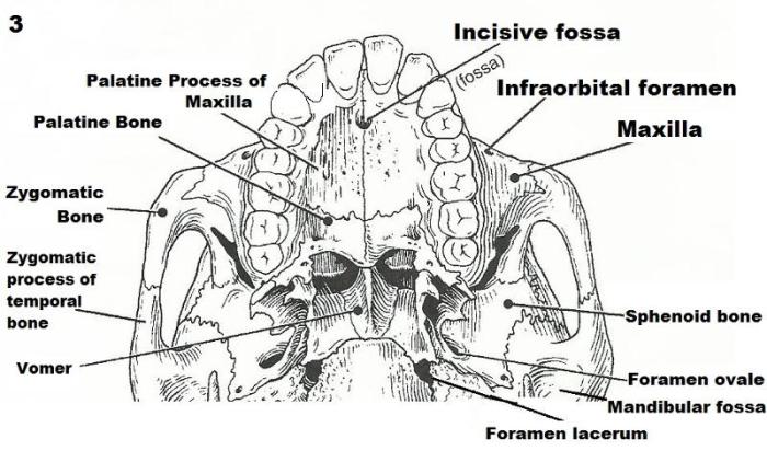
The skull can be divided into two main regions: the cranium and the facial bones.
- Cranium:The cranium, the larger and more superior portion of the skull, encloses and safeguards the brain. It consists of eight bones: the frontal bone, two parietal bones, two temporal bones, the occipital bone, the sphenoid bone, and the ethmoid bone.
- Facial Bones:The facial bones, located anteriorly and inferiorly to the cranium, form the framework of the face. They include the nasal bones, maxillae, zygomatic bones, lacrimal bones, palatine bones, inferior nasal conchae, and the mandible.
Sutures of the Skull
The bones of the skull are interconnected by a series of sutures, which are immovable joints formed by the interlocking edges of adjacent bones. These sutures not only provide strength and stability to the skull but also allow for slight growth and expansion during early development.
- Coronal Suture:Connects the frontal bone to the two parietal bones.
- Sagittal Suture:Joins the two parietal bones along the midline of the skull.
- Lambdoid Suture:Connects the occipital bone to the two parietal bones.
- Squamous Suture:Unites the temporal bone to the parietal bone.
Foramina of the Skull
The skull contains numerous foramina, which are openings that allow nerves and blood vessels to pass through. These foramina are located at various points throughout the skull and play a crucial role in the passage of essential structures.
- Foramen Magnum:A large opening at the base of the skull through which the spinal cord exits.
- Optic Foramen:Transmits the optic nerve from the eye to the brain.
- Internal Auditory Meatus:Allows the passage of auditory and vestibular nerves to the inner ear.
- Jugular Foramen:Provides a passage for the jugular vein and nerves.
Functions of the Skull
The skull serves several essential functions:
- Protection:The primary function of the skull is to protect the delicate brain from injury. Its sturdy structure acts as a protective helmet, shielding the brain from external impacts and forces.
- Support:The skull provides support and attachment for the facial muscles, allowing for a wide range of facial expressions.
- Sensation:The facial bones contain sensory receptors that enable us to perceive touch, temperature, and pain in the face.
- Resonance:The shape and structure of the skull contribute to the resonance of sound, enhancing our ability to hear and produce speech.
Nasal Cavity
The nasal cavity is a complex and vital space located within the facial bones. It serves as a passageway for air during respiration and plays a crucial role in olfaction (the sense of smell).
- Airway:The nasal cavity allows air to enter and exit the respiratory system. It filters, warms, and moistens the inhaled air, preparing it for gas exchange in the lungs.
- Olfaction:The olfactory receptors, responsible for the sense of smell, are located in the upper portion of the nasal cavity. These receptors detect and identify various odor molecules, enabling us to perceive a wide range of scents.
The Vertebral Column

The vertebral column, also known as the spine or backbone, is a flexible yet sturdy structure that forms the central axis of the human body. It consists of a series of bones called vertebrae, which are stacked one on top of the other to create a protective channel for the delicate spinal cord.
Intervertebral discs, which act as shock absorbers, are located between each vertebra.
Regions of the Vertebral Column, Exercise 9 the axial skeleton
The vertebral column is divided into five distinct regions, each with unique characteristics and functions:
- Cervical vertebrae (neck):Seven vertebrae that support the head and allow for a wide range of neck movements.
- Thoracic vertebrae (chest):Twelve vertebrae that connect to the ribs to form the rib cage, protecting the heart and lungs.
- Lumbar vertebrae (lower back):Five vertebrae that provide support for the body’s weight and enable twisting and bending movements.
- Sacrum (pelvis):Five vertebrae that are fused together to form a triangular bone, providing stability and support to the pelvis.
- Coccyx (tailbone):Four small, fused vertebrae that form the base of the spine.
Functions of the Vertebral Column
The vertebral column serves multiple essential functions for the human body:
- Support:The vertebral column provides a rigid framework that supports the body’s weight and helps maintain an upright posture.
- Protection:The vertebrae form a protective channel around the spinal cord, shielding it from injury.
- Movement:The flexible nature of the vertebral column allows for a wide range of movements, including bending, twisting, and turning.
The Rib Cage
The rib cage is a protective structure that encloses the thoracic cavity, which houses vital organs such as the heart, lungs, and major blood vessels. It consists of 12 pairs of ribs, the sternum, and costal cartilages.The ribs are curved, bony structures that extend from the thoracic vertebrae at the back to the sternum at the front.
The first seven pairs of ribs are known as true ribs, as they connect directly to the sternum via costal cartilages. The next five pairs are called false ribs, with the last two pairs being floating ribs, which do not connect to the sternum.The
sternum, also known as the breastbone, is a flat, elongated bone located in the center of the rib cage. It is connected to the ribs via the costal cartilages, forming a strong and flexible structure.The rib cage plays a crucial role in protecting the thoracic organs from external forces and injuries.
It also facilitates breathing by providing a space for the lungs to expand and contract. The rib cage muscles, such as the intercostal muscles, work together to raise and lower the ribs during inhalation and exhalation, enabling the exchange of gases in the lungs.
Movements of the Rib Cage
The rib cage undergoes various movements during respiration:
- Inhalation:During inhalation, the intercostal muscles contract, causing the ribs to move upward and outward. This increases the volume of the thoracic cavity, allowing the lungs to expand and draw in air.
- Exhalation:During exhalation, the intercostal muscles relax, allowing the ribs to return to their resting position. This decreases the volume of the thoracic cavity, pushing the air out of the lungs.
The rib cage also provides structural support to the upper body, contributing to the overall stability and posture of the individual.
Clinical Significance
The axial skeleton, comprising the skull, vertebral column, and rib cage, holds immense clinical significance in understanding and treating various disorders and injuries.
Diagnostic techniques like X-rays, CT scans, and MRIs play a crucial role in evaluating the axial skeleton. X-rays provide a two-dimensional view of bones, revealing fractures, dislocations, and other abnormalities. CT scans offer cross-sectional images, enabling detailed visualization of bone structures, soft tissues, and blood vessels.
MRIs utilize magnetic fields and radio waves to generate detailed images of soft tissues, such as the spinal cord and intervertebral discs.
Role in Surgical Procedures
The axial skeleton serves as a vital target for surgical interventions. Spinal fusion, a procedure to stabilize the vertebrae, is commonly performed to treat spinal instability, pain, and deformity. Skull repair surgeries are necessary to address fractures, birth defects, or tumor removal.
FAQ Resource
What is the primary function of the axial skeleton?
The axial skeleton’s primary function is to protect vital organs, provide structural support, and facilitate movement.
How many vertebrae are there in the vertebral column?
There are 33 vertebrae in the vertebral column, divided into five regions: cervical, thoracic, lumbar, sacral, and coccygeal.
What is the role of the rib cage in respiration?
The rib cage expands and contracts during respiration, allowing for the inhalation and exhalation of air.
